Imaging examinations and procedures
Keywords:Imaging is used to study the human body by various methods. With imaging procedures we can support surgery and partially replace vascular surgery.
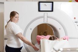
Imaging is used to study the human body using various methods. Imaging can consist of taking images, but it can also include, for example, measurements of the functions of the body, brain, or nervous system. Imaging is also used as an aid in guiding procedures and providing cancer treatments.
A physician will assess whether the examination is necessary as a whole. Imaging examinations can be used to diagnose or exclude various diseases and, if necessary, to plan effective treatment.
The most common imaging studies are X-ray examinations, MRI scans, and ultrasound scans.
With the procedures performed by imaging specialists, or radiologists, we can support surgery and partially replace vascular surgery. Radiologists can perform, for example, balloon dilatation procedures. In these procedures, they utilize imaging equipment or methods to assist them.
Diagnostic Center
How do I prepare for examinations and where do I get the results?
Examinations always require a referral. More information on the required preparations, arriving for examinations, and receiving the test results can be found here.

Units related to the service
Department of Oncology X-ray unit
In the Department of Oncology X-ray unit, we perform imaging examinations with an appointment.
Elielinaukio X-ray unit
In the Elielinaukio X-ray unit, we perform plain X-ray examinations without an appointment and other imaging examinations with an appointment.
Espoonlahti x-ray unit
In the Espoonlahti X-ray unit, we perform plain X-ray examinations without an appointment.
Honkaharju X-ray unit
In the Honkaharju X-ray unit, we perform plain X-ray examinations without an appointment and ultrasound examinations with an appointment.
Updated: 27.05.2026